The Human Brain
Functions of the Cerebral Cortex
| The Cerebral Hemispheres |
|---|
1. The cerebral hemispheres are solid structures, surrounding the cerebral ventricles that contain cerebrospinal fluid. The outside of each hemisphere is covered by a layer of cerebral cortex, about 4 mm thick, which is gray in colour due to the high density of neuronal cell bodies. The inside of each hemisphere consists of white matter - nerve fibres that run between different areas of the cortex, and connect with large nuclei in the middle of each hemisphere; the two largest nuclear masses are the basal ganglia and the thalamus. |
| 2. The surface of each hemisphere has lots of grooves - sulci - that allow the cortex to be folded and increased in surface area.
Between the sulci are the gyri: each gyrus is on the surface of the brain |
| 3.
The cortex is continuous and covers the surface of the gyri and the depths of the sulci; and on the medial and inferior surfaces of the hemispheres as well as the visible surface. The two hemispheres are connected by an enormous band of axons called the corpus callosum |
| Lobes of the Cerebral Cortex |
|---|
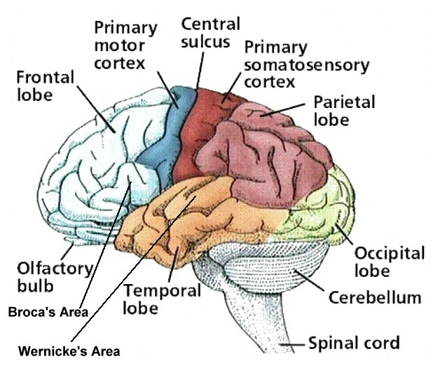
1. The central sulcus (sulcus=groove) separates the frontal lobe from the parietal lobe.
|
| 2. The frontal lobe is concerned with thinking, planning and attention. |
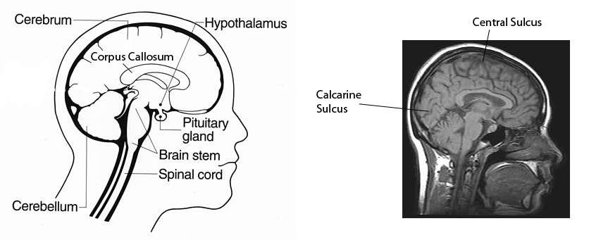
| 3.The occipital lobe is concerned with vision and visual processing. The primary visual cortex is on the medial side of the occipital lobe, surrounding the calcarine fissure. Other areas are concerned with processing of images for colour, movement, etc. |
| 4.The parietal lobe is concerned with the integration of somatosensory and visual functions; the awareness of the 3 dimensional coordinates of the surroundings of the body, and the position of limbs in that space depend on this area of association cortex. Hand-eye coordination depends on this area. Damage of this area causes astereognosis - the failure to identify or recognize objects by touch in the absence of visual or auditory inputs. Some people with damage to the parietal lobe also show neglect of the contralateral side of the body, and fail to clean or dress it. |
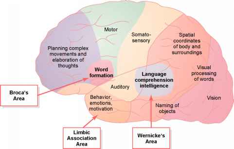
| 5. The temporal lobe is concerned with hearing, understanding speech, emotion and memory. Wernicke's area is adjacent to the primary auditory receiving area and is concerned with the understanding of speech. |
6.Broca's area is in the frontal lobe, adjacent to the primary motor area that controls muscles of the face and larynx, and is concerned with speech. |
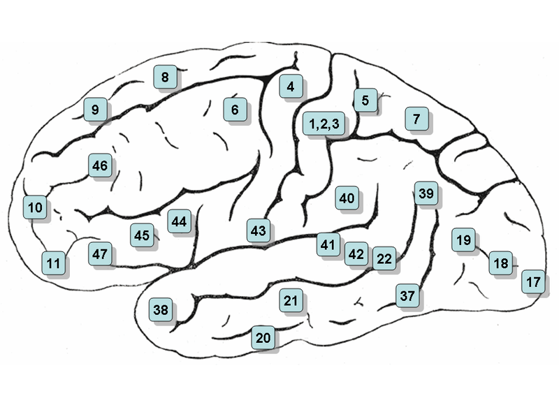
An anatomist, Brodmann, divided the cortex into different areas based on the differences in cellular structure.
Brodmann's areas 1,2 and 3 correspond to the primary somatosensory receiving area and area 4 is the motor cortex.
Area 6 consists of the pre-motor cortex and supplementary motor area (M2), and areas 17, 18 and 19 are concerned with different aspects of visual processing.
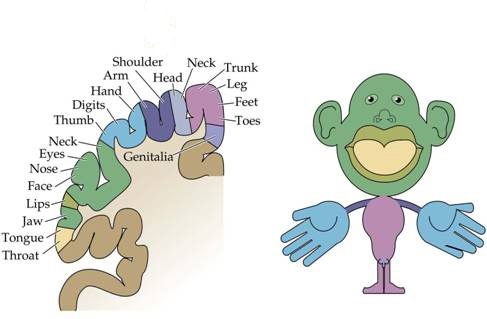
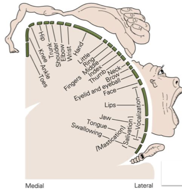
On the left is a diagram of a sagittal section of the somatosensory cortex. On the right is a drawing showing the disporportionate amount of cortex given to the face, lips and hands.
| Somatosensory Cortex |
|---|
| 1. There is a general rule that one cerebral hemisphere handles sensory inputs from the opposite side of the body. |
| 2. Sensory inputs to the cortex arise from the thalamic nuclei, and there is a topographic map of one half of the body's surface in the contralateral thalamus and somatosensory cortex. |
| 3. Areas of the body surface with greatest two-point discrimination (i.e. densest innervation) have the largest areas of somatosensory cortex. The somatotopic map of the body surface on the cortex is therefore distorted, depending on the density of innervation of the skin, and is called the homonculus. |
| 4. Areas 1, 2 and 3 are strips of cortex each dealing with a different type of sensory input from the skin, muscles and joints. |
| 5. |
| 6. Damage to the sensory cortex results in decreased sensory thresholds, an inability to discriminate the properties of tactile stimuli or to identify objects by touch. |
| The Motor Cortex |
|---|
| The motor cortex on the pre-central gyrus contains an orderly map of all the possible movements of the opposite side of the body arranged in order; electrical or magnetic stimulation of each small area of motor cortex causes contractions of groups of muscles responsible for a specific movement. |
| Stimulation of each area within the somatotopic map induces contractions of relevant muscle groups rather than of individual muscles. |
| Electrical stimulation within a large area in the middle of the motor cortex is concerned with the control of hand muscles. The thumb has a particularly wide range of movements including opposition - the ability to direct it to touch every fingertip and pick up objects. |
| The muscles of the face also have a disproportionate representation on the cortex; this is partly associated with the muscles of facial expression, but also with the muscles of the jaw and others involved in swallowing. |
| The corticospinal tract is the executive pathway in the control of voluntary movements, and connects the motor cortex with alpha motoneurones. |
| The motor cortex and cortico-spinal tract are particularly concerned with producing fine, precise movements. Lesions of the motor cortex (such as the surgical ablation of one area) lead to paralysis or a loss of power in the muscles affected. In time, these muscles can regain some movement, but the movements are always coarse in comparison. |
| Sometimes the corticospinal tract (pyramidal tract) is called the 'upper motoneurone' because it controls the activity of the 'lower motoneurone' (i.e. the alpha motoneurones). |
| The corticospinal tract is also called the pyramidal tract because it crosses the midline in the pyramids of the medulla. The term 'extrapyramidal' refers to pathways that can cause coarser movements, mediated by other pathways. |
The association areas are adjacent to the primary receiving areas, e.g. the parietal lobe uses information from the somatosensory and visual cortices to guide the limbs, fingers and eye movements and generate complex movements requiring inputs from skin and eyes. Lesions of these areas leads to a failure to integrate sensory information. Areas behind the postcentral gyrus integrate visual and spatial inputs and are involved in perceiving an awareness of trajectories of moving objects. Unsurprisingly proprioception (awareness of the position of body parts in space) is also represented in this area..
The somatosensory association cortex (Brodmann's areas 5 and 7) receives inputs from SI and SII and lies directly behind the sensory cortex in the superior parietal lobes. Damage to this area causes tactile agnosia, an inability to recognize objects even though the objects can be felt. lesions of the parietal lobe also cause a neglect syndrome in which the patient is unaware of the opposite side of the body, and does not wash or clothe it.